High Full Well cameras bring pathology to the operating room.
Pathology will move into the operating room (OR). Acquiring histology information from biopsies in 2 minutes or directly from the patient without a biopsy will become feasible in the coming years.
This will all be made possible by looking directly at the cellular level through innovative Full-Field OCT and Dynamic Cell Imaging.
A few of the benefits of this development are among others are:
- Maximize localization of the relevant tissue for cancer diagnostics and surgery.
- Better determination of the essential tissue for diagnostics. This reduces the repeat biopsy rate, that is sometimes as high as 30%.
- Reduction of surgery time and validation of complete focal ablation.
With direct diagnostic information from the OR and faster diagnostics turn-around times resulting in lowered costs and increased patient satisfaction.
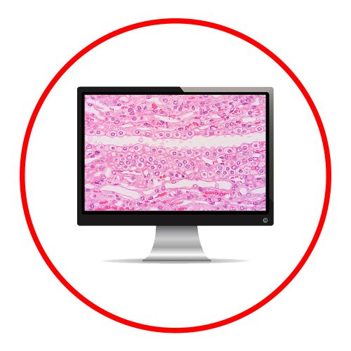
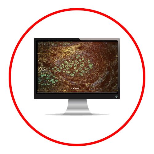
The ability to look instantly inside transparent matter with Full Field OCT and Dynamic Cell Imaging microscopy (LLTech) is fueled by Teledyne Adimec High Full Well camera technology. This new imaging method creates the ability to quickly recognize & diagnose pathology information without the need to prepare the tissue through slicing & staining. Teledyne Adimec developed the High Full Well camera technology in the CAReIOCA FP7 project. This technology is now available in our Quartz Q-2HFW camera. With a Full Well depth over two million electrons per pixel (e.g. high SNR), 2 Mpx imaging resolution and frame speed up to 550 fps (full frame), this camera allows to observe fast changing small contrast variations in bright scenes under conditions not possible before.
 English
 日本語
日本語 简体中文
简体中文