Adimec (Netherlands) together with five partners, as part of the CAReIOCA consortium are developing & validating a high-resolution high-speed imaging based medical device to perform non-invasive optical biopsy for cancer assessment. Adimec contributes with a specific high-performance CMOS CoaXPress camera.
The project was started at the beginning of 2013, results were released in June 2014 and now some additional results were published in the third newsletter. The key achievements in the second half of 2014 include:
- The integration of the CMOSIS sensor into an ADIMEC camera
- Leiden University Medical Center and Institut Gustave Roussy completed the atlases containing comparative FFOCT and histological images of pathological and healthy biopsies, respectively taken from patients suspected of breast or head and neck cancer
- LLTech is developing the two FFOCT imaging device prototypes and preparing to deliver a preliminary version of the endoscope in December 2014
The goal of CAReIOCA is to provide pathologists and/or surgeons non-invasive optical imaging at the cellular level within the human body in real time. The optical technology used in the end system is based on Full Field Optical Coherence Tomography (FFOCT), a technique that enables volumetric image capture on semi-transparent tissue at micron resolution in 3D. The technology intends to assist in diagnosis, particularly of cancer, in skin, breast, prostate, brain etc, as well as quality control of biopsies.
High-speed, extreme low shot noise CMOS CoaXPress camera developed
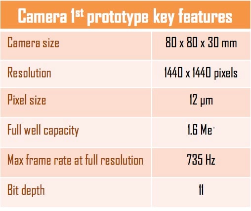
The first camera prototype was assembled by ADIMEC and delivered to LL Tech at the beginning of July. The image sensors were characterized by CMOSIS and then transferred to Adimec to embed the image sensor in a camera platform with sufficient high-speed precision imaging and interfacing in order to plug in easily in the LL Tech FFOCT devices. The camera utilizes a CoaXPress interface, which is the serial communication standard best, suited for high-speed heavy data transfer.
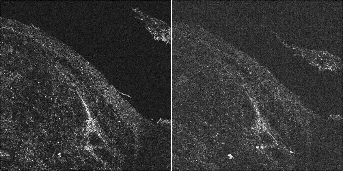
Tests and prototyping continue at CMOSIS and ADIMEC. LLTech has integrated the first camera prototype on their commercial LightCT scanner and some images have been recorded in order to appreciate the benefits of the exceptionally high full well capacity (e.g. extremely low shot noise level). As an example, comparative images of a skin sample are shown below. The left image was obtained with a CAReIOCA camera prototype whose full well capacity (FWC) was measured at 1.6 million electrons (1600 kel) whereas the right image is taken with the camera usually embedded in LightCT scanners, of 0.2 million electrons (200 kel) FWC. Both images required only one accumulation. These images show that the signal to noise ratio (SNR) is at least two times enhanced with the CAReIOCA camera. A 3-fold gain is expected on the SNR with further optimization of the camera parameters. The new camera will also increase the acquisition speed by a factor of 5.

Image from the CAREiOCA camera Image from typical camera
The new camera was demonstrated at the Vision Show in Stuttgart, Germany and the ITE show in Yokohama, Japan. At the Photonics West 2015 exhibition the new camera will be presented in the context of the application it is designed for, which means it is integrated in a LLTech FFOCT microscope in the Adimec/LL Tech booth. Clinical results of this new microscope technology will be presented at several sessions during the BiOS conferences at Photonics West 2015.
Completion of the clinical atlases
The clinical partners in this project are Leiden University Medical Center (LUMC) in Leiden, the Netherlands and Gustave Roussy Institute (IGR) in Villejuif (Paris), France. LUMC perform studies on breast cancer while IGR is focused on studying head & neck cancer.
In the summer of 2014, LUMC and IGR completed the clinical atlases, which are databases of comparative FFOCT/histology images designed to develop educational and diagnosis capabilities based on FFOCT images. They contain typical images of non-pathological and pathological tissues organized by anatomical sites and tissue abnormality types. FFOCT images are displayed in both positive and negative contrast, next to the corresponding histological slide, and include annotations pointing out morphological features, interesting observations and diagnosis criteria.
FFOCT imaging device prototypes
LLTech completed the design of the two FFOCT devices. The design of the high-speed high-resolution microscope is an adaption of the existing commercial LightCT scanner to the application of large-scale tumor margin observation. The optical system remains unchanged except for the integration of the new camera and the main developments concern the sample holder, the associated travel mechanics and the software.
As for the FFOCT contact endoscope, the design is unprecedented and based on an original optical system of tandem interferometry that guarantees an easy replacement of the probe and a compact device. The choice of components is mainly motivated by image quality and compactness requirements.
This project has received funding from the European Union Seventh Framework Program FP7-ICT-2011-8 under grant agreement number 318729.
To get more details on the CAReIOCA project, click here.
 English
 日本語
日本語 简体中文
简体中文


