In many countries there is a demographic shift to older age groups resulting in increased incidences of cancer and many other diseases. This means an increased number of patients and samples while the number of skilled pathologists is not growing at the same rate. There is also a desire, especially with the assessment of cancer, to decrease the time for diagnosis to improve treatment results.
Pathology labs need to continuously increase output and efficiency while also improving quality. Automated digital microscopes, digital slide scanners, and other digital pathology systems allow for digitizing of the images pathologists previously viewed manually and open up new ways of operating. It reduces the time required by pathologists, allows for expert opinions remotely (telepathology) without shipping slides, enables more efficient data storage, and other benefits.
The Adimec 12 Megapixel cameras (Q-12A65 and Q-12A180) offer the high-resolution, high frame speed combination to provide throughput and image detail requirements of these systems. The exceptional image quality of these cameras delivers the necessary input data for further analysis. Functionality such as region of interest (ROI) allows for portions of the image to be checked at a lower bandwidth during settings adjustments to quickly and automatically identify the proper sharpness settings before full resolution images are obtained. Additional features for image tracking are possible.
FFOCT will provide a complementary technique to histology as it can be used during surgical procedures. The image below shows a comparison of FFOCT and histology. The image was provided by LLTech.
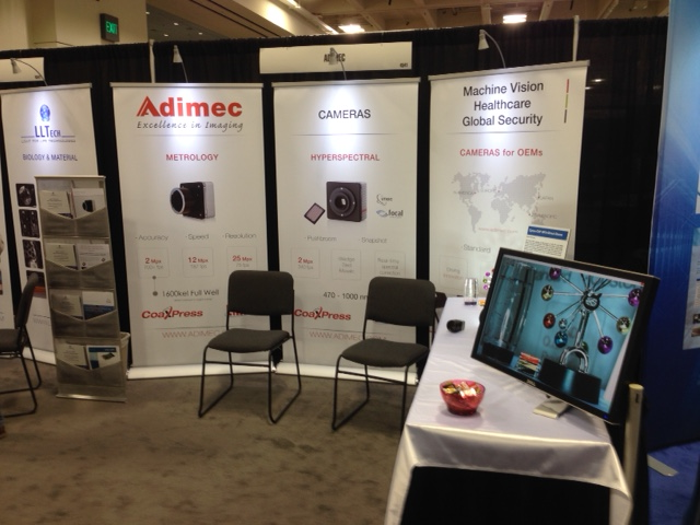
To learn more about Adimec’s extreme full well camera or any of our cameras, please visit us at booth #4541 at Photonics West this week.
 日本語
日本語 English
 简体中文
简体中文


